Introduction
1.1 Background on the Subject Matter
The brain is a complex organ in the human body, and its structure varies from one individual to the other. The human brain has many components, including grey matter, areas in the brain mainly consisting of synapses, neuronal cell bodies, and glial cells (Bigbee, 2022). Grey matter is vital in many cognitive functions, including controlling people’s attention, memory, and perception (Zidan et al., 2022). Like other athletes in contact sports, Amateur boxers are exposed to multiple potential risks of repetitive head impacts, which can significantly have long-term impacts on the brain health of amateur boxers (Kurtoğlu et al., 2023). Continuous head trauma, like the cumulative effects of boxing, may result in functional and structural changes in the brain, which could reduce the grey matter volume (Aranha et al., 2022). Zidan et al. (2022) explain that determining the exact volume of grey matter in the brain of an amateur boxer requires a comprehensive brain imaging study like a high-resolution MRI scan, and advanced image analysis techniques should follow this.
One common risk amateur boxers are constantly exposed to is mild traumatic brain injury (mTBI) (Zidan et al., 2022). MTBI refers to a brain injury due to a jolt or blows to the head, thus leading to temporary dysfunction of brain functions. This condition is mainly prevalent among athletes who participate in the contact sports like boxing and among individuals in the military (Bernick & Banks, 2020). According to Zidan et al. (2022), approximately 42 million people globally sustain mild traumatic brain injury (mTBI) annually, which shows that the condition significantly impacts public health. There have been inconsistencies in the evidence on the impacts of mTBI, particularly in amateur boxers, as many studies have produced conflicting results. Some research shows that amateur boxers who sustain mTBI have a deterioration in cognitive abilities (Bernick & Banks, 2020). This further proves that repetitive head trauma may harm brain health and cognitive functions. Even without overt cognitive impairment, biomarker analysis of cerebrospinal fluid (CSF) has shown substantial evidence of traumatic axonal injury in amateur boxers. However, some research has cast doubt on the link between amateur boxing and long-term brain damage (Zidan et al., 2022). This inconsistency in results illustrates the difficulty of researching the long-term effects of mild traumatic brain injury.
Zidan et al. (2022) state that standard CT and MRI scans often fail to address the subtle structural alterations typical of mTBI, such as contusions and microhemorrhages. Diffuse axonal damage, which may be seen with diffusion tensor imaging (DTI), and cortical thinning, which can be measured using high-resolution (HR) structural magnetic resonance imaging, are two examples of more subtle changes that can occur. The thalamus, caudate nucleus, and putamen are all brain regions vulnerable to damage from repeated head trauma. These areas are a part of the basal ganglia, responsible for various activities, including movement, cognition, and emotion control (Zidan et al., 2022). The years spent by the professional boxer during training have been linked to a decrease in volume in these subcortical gray matter areas. This implies that experienced boxers who have been in the sport for a long and have been exposed to many boxing fights are more likely to have brain volumes that are smaller in the caudate nucleus, the putamen, and the thalamus (Zidan et al., 2022).
1.2 Research Question
Zidan et al. (2022) aimed to evaluate the variances in the volume of anatomical brain structures between the amateur boxers and the control subjects, focusing on the impacts of this condition on the grey matter. Specifically, their research focused on answering the question, “How do the thalamus, caudate nucleus, and putamen volumes change in amateur boxers who experience frequent head trauma compared to age-matched healthy controls?”
1.3 The Hypothesis and Predictions of the Authors
Zidan et al. (2022) hypothesized that amateur boxing would harm the volumes of deep grey matter structures compared to non-athletes of the same age. The authors further hypothesized that the repetitive blows on the heads of amateur boxers are likely to cause significant attenuation of the volume of some areas of the brain’s deep grey matter. The ultimate notion is that the physical demands and repetitive head hits in amateur boxing contribute to the development of mild traumatic brain injuries (mTBI), even if cognitive problems may not be immediately apparent. The authors further predicted that there would be a sizeable distinction in the quantities of deep grey matter between amateur boxers and age-matched healthy controls (Zidan et al., 2022). According to their prediction, the caudate nucleus, thalamus, and possibly the putamen will have smaller sizes in amateur boxers. This prediction is based on research linking mild traumatic brain damage (mTBI) to long-term neurodegenerative cascades and on studies of professional fighters demonstrating volume reduction in these regions following repeated head trauma.
Methods
This study aimed to effectively examine the correlation between amateur boxing and changes in brain volume and cognitive ability by comparing boxers and non-boxers. The study was conducted following the ethical standards set by the institutional ethics board, together with informed consent from all the parties who participated in this study. 19 male amateur boxers from a local Olympic base and 19 healthy male control subjects without a history of participating in boxing or any other contact sports participated in the study. The participants were drawn from a previously published sample. The researchers used age and IQ to effectively ensure equality between the two groups. Participants were not allowed to have a history of substance abuse, arterial hypertension, neurological or mental disorders, metabolic diseases, or any other abnormalities in the brain that would confound the study’s results. The brainpower of the participants was measured through the use of a series of neuropsychological exams. Memory performance was measured using the Verbal Learning and Memory Test (VLMT), alertness, planning, and memory were evaluated using the “alertness” subtest from the “test of attentional performance” (TAPAL), and IQ was measured using the Revised Hamburg Wechsler Intelligence Scale (HAWIE-R).
As part of the anthropometric evaluation, the weight and BMI of the participants in this study were evaluated. An MRI was obtained by scanning each patient in a 3T MR scanner. The MRI included several sequences, including axial time-of-flight (TOF) MR angiography, coronal T2*-weighted imaging, transverse dual spin-echo imaging, and 3D sagittal magnetization-prepared rapid acquisition of gradient echo (MPRAGE). Both the structure and function of the brain were revealed by these scans. After collecting 3D MPRAGE images, the researchers ran them through the volBrain processing pipeline. Preprocessing on numerous fronts was necessary for this, including standardizing intensity, registering to a reference brain template, smoothing out inhomogeneities, and denoising. The researchers further used different techniques, including nonlocal intracranial cavity extraction (NICE), tissue classification, nonlocal hemisphere segmentation (NABS), and nonlocal subcortical structure segmentation, to effectively make educated guesses regarding the brain’s volumes of the grey matter. All statistical evaluations and analyses were performed in SPSS.
The researchers employed descriptive statistics to compile a broad overview of our subjects. Brain volumes were compared between boxers and controls using Student’s t-tests (total intracranial volume [TIV], gray matter [GM], white matter [WM], and normalized brain volume [NBV]). Analyses of correlation analyzed the connection between body mass index and brain volume. Deep gray matter structures were compared between groups using multivariate variance analysis and covariance analysis, controlling for age and body mass index. The regional grey matter volumes of the athletes were analyzed using a linear regression model to determine if there was a correlation between the number of fights they had and how long they had been boxing.
Results
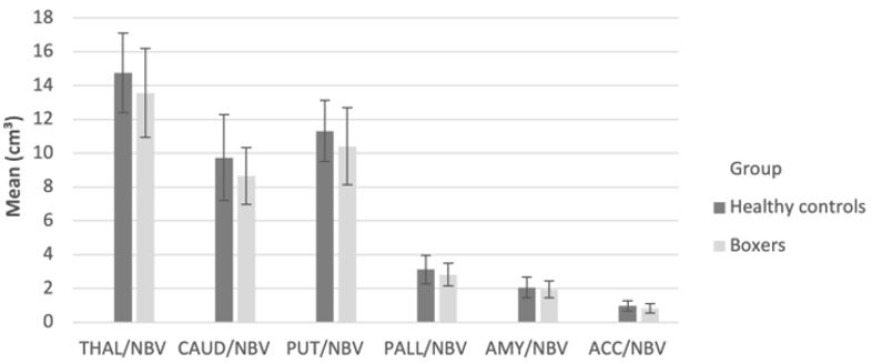
Figure 1: Analysis of boxer’s deep grey matter structures
Figure 1 was generated using many statistical methods to compare the volumes of deep grey matter structures in a group of boxers and a healthy control group (HC). The researchers compared the volume of the grey matter components between the two groups through a multivariate analysis of variance (MANOVA) with body mass index (BMI) as a factor. Covariance (ANCOVA) analysis was used to adjust the data for the potential influences of age effectively. This analysis involved considering the ages of the participants as a covariate to limit the effect of age on the measurements of grey matter thickness. The researchers further used linear regression studies to identify the number of fights a boxer had, the number of years they have taken in the sports, and the correlation to the brain sizes in various regions.
The bar graphs in Figure 1 depict the volume of the deep grey matter structure and represent the range of possible values for the measurements. Using the data presented in the figure in the context of the study’s research question, one can closely look for discrepancies in the volumes of the deep grey matter structure between the boxers and the controls. The error bars can determine the variation in the measurements and the statistical significance of the differences between the two groups. The numbers may also shed light on the correlation between boxers’ regional brain sizes and the total number of fights and years spent in the sport. The linear regression analysis shows that these connections help illuminate the possible effects of boxing training on various brain regions.
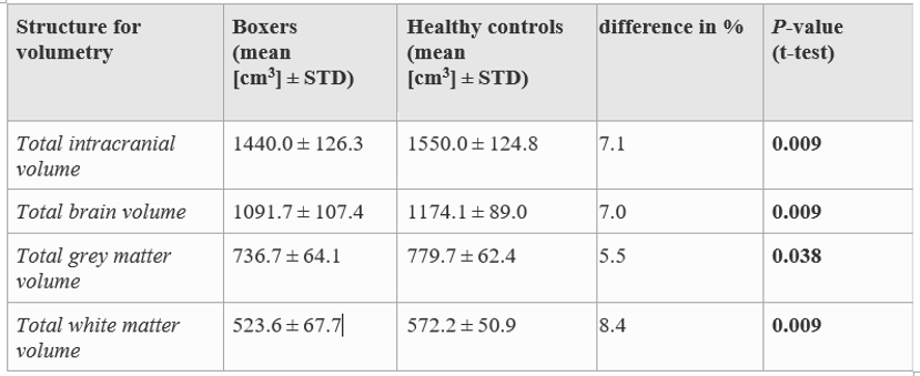
Figure 2: Quantitative analyses of the brain, white matter, and grey matter volumes
Figure 2 presents data illustrating the findings of a qualitative analysis of the brain’s grey matter and white matter volumes among amateur boxers and a group of healthy control across a range of volumetric metrics. The analysis quantifies the volume of the brain, white matter, and grey matter in addition to the intracranial volume (TIV). Both the mean values and standard deviations (STD), given in cubic centimeters (cm3), were provided for all the volumetric measurements. The “difference in%” shows the proportional disparity between the Boxers and Healthy norms. The study further examined the relationship between body mass index and the volumetric data. The data shows that there is a positive relationship between body mass index and TIV (r = 0.42, p = 0.009), TBV (r = 0.46, p = 0.003), and TWMV (r = 0.38, p = 0.019). The connection between body mass index and TGMV was weak (r = 0.06, p = 0.721). The results presented in Figure 2 reveal that total intracranial, brain, grey matter, and white matter volumes were all lower in Boxers than in Healthy controls. Total intracranial volume, brain volume, and white matter volume were positively correlated with body mass index, while total grey matter volume was not correlated with body mass.
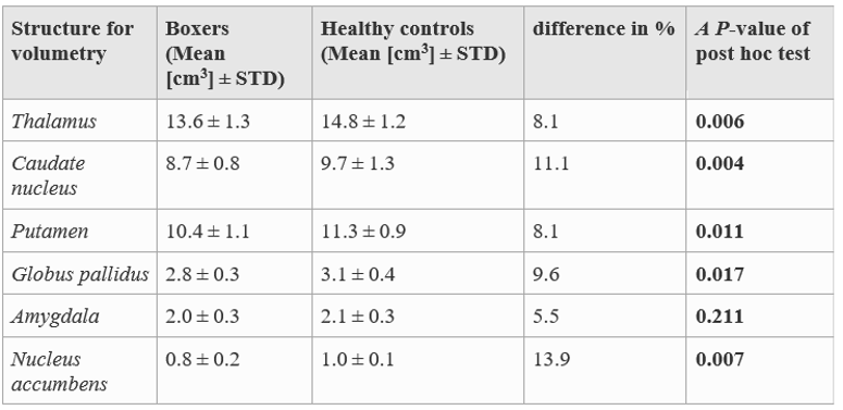
Figure 3: Quantitative volumetric of the structures of grey matter at different depths
Figure 3, above, is a representation of the data obtained from a multivariate investigation of deep grey matter structural volumes in boxers and healthy controls. The caudate nucleus, thalamus, nucleus accumbens, putamen, amygdala, and globus pallidus were all measured for their volumes by scientists. The data includes the average volume in cubic centimeters (cm3) and the standard deviation (STD) for both boxers and healthy controls for each structure. In addition, the data displays the percentage volume difference between the two groups and the p-values from post hoc tests that assess the significance of the volume differences. The researchers also used linear regression analysis to examine how the years spent boxing correlate with the sizes of various structures. The data show that the longer one engages in boxing, the smaller the right thalamus becomes (= -0.811, p = 0.049). A negative correlation was also seen with the right globus pallidus (= -0.765, p = 0.066). There were no significant connections between the number of fights and the remaining volumetric measurements in boxers, and no significant impacts were detected for the predictor factors.
Discussion
4.1 Authors’ Conclusion
The findings of this study reveal that the results obtained are consistent with the proposed framework. The authors postulated that amateur boxers, compared to healthy controls, would have different deep grey matter architectures, notably a decreased volume in specific brain regions. Results showed amateur boxers had smaller thalamic, caudate, putamen, globus pallidus, and nucleus accumbens volumes than healthy controls. Zidan et al. (2022) explain that the most considerable differences in the volume of grey matter in the brain of the participants were mainly found in the nucleus accumbens, followed by the caudate nucleus and globus pallidus. Notably, the findings obtained in this study are closely related to the previous studies on moderate to severe traumatic brain injury (TBI) and the neurodegenerative diseases that are associated with the loss of GM volume. Zidan et al. (2022) explain that the thalamus, which plays a vital role in relaying sensory information, reduced the volume of grey matter among the boxers in this study. The results of this study were found to be in agreement with those of biomechanical studies, which found that the thalamus experiences greater shearing stress in the event of head impacts.
The study demonstrates that even with repeated exposure, amateur boxing can induce long-term damage to the brain and structural abnormalities in specific brain regions. Previous research on traumatic brain injury (TBI) and neurodegenerative illnesses has found volume reductions in deep grey matter tracts. The findings highlight the need to think about the effects of these behaviors on mental well-being. The writers make a strong argument for their analysis of the data. They discuss how the study’s limitations, like its small sample size and absence of longitudinal data, weaken its overall findings and conclusions. The authors also acknowledge the possibility that the different backgrounds, environments, and genetic profiles between the boxers and controls contribute to the observed discrepancies.
4.2 Personal Conclusion
The authors make reasonable inferences from the obtained data. However, the study has some flaws that should be considered. Given the limited sample size and the lack of longitudinal data, it is crucial to approach the findings cautiously. Longitudinal data would have helped researchers understand how brain shrinkage and boxing exposure changed. Testing individuals’ brain architecture and neuropsychological performance many times would have helped catch any changes and determine whether they correlate with boxing-related injuries. Furthermore, the study’s small sample size diminishes its statistical power and potentially restricts the generalizability of its findings. A bigger sample size would have allowed for the detection of more nuanced relationships between changes in brain volume and behavioral measures. A larger sample size also allowed for subgroup studies, such as the impact of weight class or time spent boxing on brain sizes, which could shed light on the significance of various aspects of brain health.
The study also had several holes, such as not evaluating when possible injuries occurred among amateur boxers. The study’s assumption that boxers sustain cumulative injuries throughout their careers raises the possibility of bias in the resulting data. It would have been more informative to collect data on the frequency and timing of bouts and correlate them with changes in brain structures. More information about the acute and long-term effects of mTBI in boxing may have been gained from a better understanding of the temporal relationship between injuries and brain changes.
Future research should use a longitudinal approach with a bigger sample size to bolster the study. The association between boxing exposure, brain atrophy, and neuropsychological results can be understood effectively through longitudinal evaluations. More people in the sample would improve the study’s statistical power and make its results more generalizable. In order to further establish the temporal association between injuries and brain alterations, it would be helpful to collect specific information on the timing and frequency of head hits connected to boxing.
References
Aranha, M. R., Coutinho, A. M., de Godoy Carneiro, C., Pastorello, B. F., Studart-Neto, A., Guariglia, C. C., & Leite, C. C. (2022). Brain glucose metabolism and gray matter volume in evaluating chronic traumatic encephalopathy in retired professional soccer players: a cross-sectional [18F] FDG-PET/MRI study.
Bernick, C., & Banks, S. (2020). What boxing tells us about repetitive head trauma and the brain. Alzheimer’s research & therapy, 5(3), 1–6.
Bigbee, J. W. (2022). Cells of the Central Nervous System: An Overview of Their Structure and Function. Glycobiology of the Nervous System, 41-64.
Kurtoğlu, E., Payas, A., Düz, S., Arık, M., Uçar, I., Tokmak, T. T., … & Unur, E. (2023). Analysis of changes in brain morphological structure of taekwondo athletes by diffusion tensor imaging. Journal of Chemical Neuroanatomy, 129, 102250.
Zidan, M., Jesser, J., Herweh, C., Jost, J., Heiland, S., Meyding-Lamadé, U., … & Haehnel, S. (2023). Deep grey matter volume is reduced in amateur boxers compared to healthy age-matched controls. Clinical Neuroradiology, 33(2), 475-482.
 write
write