The chosen audience for learning the interactions between the muscular, nervous, and skeletal systems are medical patients. In particular, the activities that can help medical patients understand the functions of the skeletal, muscle, and nerve systems include bicep curls, squats, shoulder presses, walking, and neck rotation because they enable people to make informed choices about their health or decide what types of activities they can engage in more effectively.
Activities
Bicep Curl
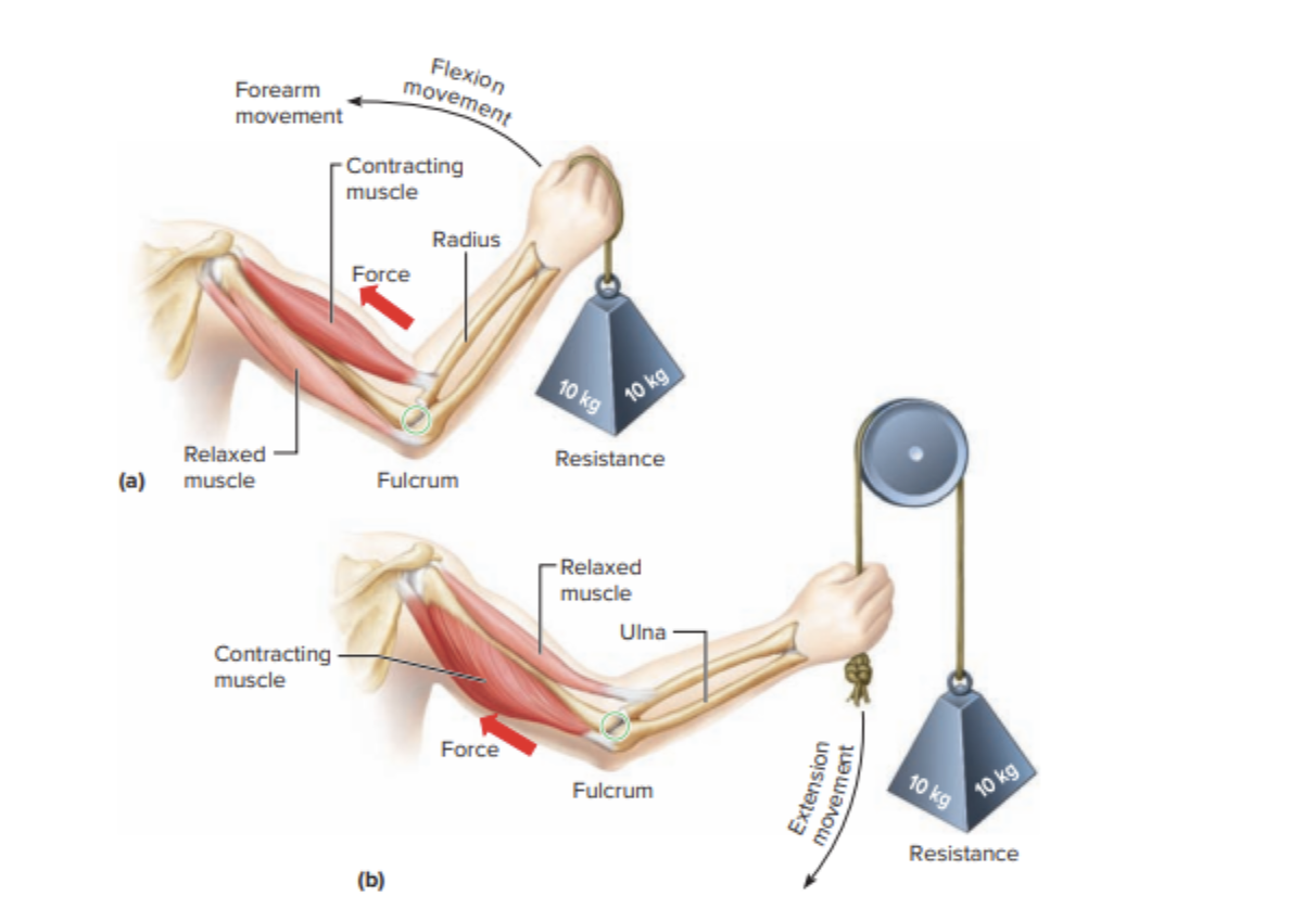
Elbow flexion is the contributing movement of the bicep curl. For this exercise, position yourself with the dumbbell between your thighs, holding it in an overhand grip such that you have to support its weight on one hand. Flex the elbow and pull the dumbbell towards the shoulder (See Figure 1). The elbow joint complex consists of different articulations formed by the forearm and arm bones, including the ulna and humeral joints (Islam et al., 2020, p. 95). Elbow flexing is accomplished at the elbow joint, which is responsible for promoting movement. The movers in the bicep curl are the muscles of the upper limb, which are attached to bones that then move with the help of the nerves.

Figure 1. Bicep curl. Adapted from “Hole’s essentials of human anatomy & physiology” by Charles & Cynthia, 2020 p. 205, copyright 2020 by McGraw Hill.
Squats
The knee and hip joints contract for forward flexion of the knees while stretching them during backward extensions is essential for squats. When doing one squat, position your legs wider than they should be, bring down your body by bending the knees, and go back to the initial point. The bones engaged in this articulation are located in the lower limb, including the femur, tibia, and fibula (Charles & Cynthia, 2020, p. 172). The knee and hip joints are responsible for flexion and extension of the lower limb. Here, the primary muscle groups involved include the muscles of the front thigh, with some assistance from the lower back and back thigh muscles.
Shoulder Press
In the shoulder press, there is a joint movement of the arm at the level of one’s joint. The dumbbells should be on the shoulders with the palms facing forward. The dumbbells should be lifted over toward the head and then back down. The scapula, clavicle, and humerus are the bones that participate in this movement (Charles & Cynthia, 2020, p. 166). A shoulder joint permits outward and inward movement of the arm through the connections of bones and the muscles that form the joint. In this activity, the shoulder muscle is a prime mover muscle with the help of other arm muscles.
Walking
The flexing and extending of the hips occurs at the knee joint during walking. To do this move, go from one side to another as you bring your hands towards the ground, going in opposite directions with each leg. The bones affected by this movement include the femur, hip, tibia, knee, fibula, ankle bone, and foot (Charles & Cynthia, 2020, p. 172). Movement of flexion and extension takes place at the hip joint. At the same time, as it permits all movements, the knee joint allows for flexion and extension (See Figure 2). In this function (walking), the gluteal muscles are the prime movers, with a contribution from muscles of the front and back thigh as secondary muscles that aid the walking.

Figure 2. Bicep curl. Adapted from “Hole’s essentials of human anatomy & physiology” by Charles & Cynthia, 2020 p. 205, copyright 2020 by McGraw Hill
Neck Rotation
As for neck rotation, it implies the twisting of backbones located at the neck region called the cervical vertebra. To do this movement, sit or stand with an upright back and slowly look from one side to the other. A reduced model was verified using experimental data from in vitro tests that assessed the range of motion of the seven cervical vertebras in terms of rotation, lateral bending, and flexion extension. (Silva et al., 2022, p. 1). The pivot joint between the first two cervical vertebrae provides neck rotation. Thus, the primary propulsion muscles in play are the neck muscles, which contract to cause rotational movement.
Nervous System Process
The movement signals originate from inputs that are integrated, initiated, and coordinated by the brain, spinal cord, and the nerves in the rest of the body. It has long been believed that motor information influences movement through descending nerves originating from the brain (Karadimas et al., 2019, p. 1). When the brain decides to move, nerve impulses flowing from the brain pass through the spinal cord and transmit into muscles via the nerves (See Figure 3). Nerve impulses move longitudinally along the neurons, resulting in pathways able to conduct these signals. Hence, the transmission of the signal between nerves and muscles occurs due to chemical messengers that cause precise coordination in contractions for movement.

Figure 3. Bicep curl. Adapted from “Hole’s essentials of human anatomy & physiology” by Charles & Cynthia, 2020 p. 205, copyright 2020 by McGraw Hill.
In conclusion, the communication between the skeletal, muscular, and neurological systems is critical for human movement. The bicep curl brings the radius, ulna, and humerus bones to work in the elbow joint, with the arm’s prime mover being the muscles. Squats entail flexion and extension of the joints at the knee and hip, using the quadriceps, hamstring muscles, and muscles of the gluteal region. Lastly, during walking, hip flexion and extension vary. At the same time, the knee and ankle joints also undergo cyclic activity, with most of these movements supported by thigh and lower back muscles.
References
Charles. J. W & Cynthia. P. C., (2020). Hole’s essentials of human anatomy and physiology. McGraw-Hill Education.
Islam, S. U., Glover, A., MacFarlane, R. J., Mehta, N., & Waseem, M. (2020). The anatomy and biomechanics of the elbow. The Open Orthopaedics Journal, 14(1), 95–99. https://doi.org/10.2174/1874325002014010095
Silva, A. J. C., Alves de Sousa, R. J., Fernandes, F. A. O., Ptak, M., & Parente, M. P. L. (2022). Development of a finite element model of the cervical spine and validation of a functional spinal unit. Applied Sciences, 12(21), 1-15. https://doi.org/10.3390/app122111295
Karadimas, S. K., Satkunendrarajah, K., Laliberte, A. M., Ringuette, D., Weisspapir, I., Li, L., Gosgnach, S., & Fehlings, M. G. (2019). Sensory cortical control of movement. Nature Neuroscience. 1-24 https://doi.org/10.1038/s41593-019-0536-7
 write
write