Background
Hard deposits formed of minerals and salts in the kidney are called kidney stones, also known as nephrolithiasis. In the United States, one in every eleven people has kidney stones. Men experience kidney stones at a rate of about 80%. This happens most frequently between the ages of 20 – 30 (Mayo Clinic, 2022). Kidney stones are more common in women and do so later in life. Several variables, including genetics, the environment, a person’s food, and lifestyle, can cause kidney stones to grow. A possible connection has been proposed between kidney stone production and eating more high fructose corn syrup, fast food, and climate variation. While passing nephrolithiasis can be excruciatingly painful, they rarely have long-term consequences (Siljeholm, 2017). Based on the situation, one might need to take painkillers and drink water to get rid of a kidney stone. Surgery is occasionally necessary. While passing nephrolithiasis can be brutally painful, they typically do not result in permanent damage if caught early enough (Puckett, 2016). Depending on the needs, one might require painkillers and water to get rid of a kidney stone. Surgery might be required in other situations, such as when stones obstruct the urinary tract, are connected to inflammation, or cause issues. We will discuss kidney stones’ etiology, prevention, and treatment and the normal anatomy and physiology of the impacted primary body systems.
Normal anatomy of effective body systems affected.
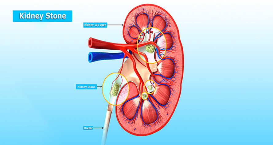
The body’s urinary system is the s drainage system for getting rid of extra water and waste in the urinary bladder; kidneys, ureters, and urethra are all parts of the urinary system. The kidneys are a pair of bean-shaped, fist-sized organs situated immediately below the rib cage in the middle of the back. The right kidney is lower than the left around the liver—the fascia, back muscles, and fat shield the kidneys. Urine travels through the ureter from the kidney to the bladder, where it is stored until it is expelled from the body by the urethra (Siljeholm, 2017). Typically, a person has two kidneys. Here is the kidney diagram showing where the kidney stone is affected.
Normal physiology of the body system is affected.
The urinary system’s primary functions are to assist in controlling the water balance and eradicate dangerous toxins from the blood. The kidneys produce urine to filter the blood. The two kidneys process about 200 quarts of blood to create 1 to 2 quarts of urine. Waste and potentially dangerous substances can be found in urine. The kidneys excrete waste and extracellular fluid (urine) through the ureters. Each kidney has its ureter, making a total of two. Peristalsis is used by the ureters, lining-covered muscular tubes, to push and drain urine into the bladder. Indications are frequently absent until a kidney stone passes through or enters one of the ureters. The tubes that link the bladder and kidneys are known as ureters. A little stone may move out of the way, hurting little or nothing. The urinary system may become obstructed by a bigger stone. A kidney stone can block urine flow, resulting in excruciating discomfort and bleeding (Siljeholm, 2017).
Mechanism of pathophysiology
High concentrations of specific minerals in urine are what produce kidney stones. Urine contains calcium, oxalate, and phosphorus. For certain people, certain meals can result in kidney stones. Assume you have a situation that alters the thresholds of intoxicants in your urine, a family medical hx of kidney stones, recurrent or reoccurring urinary tract impurities, urinary tract blockage, digestive issues, or repeat UTIs. The likelihood of developing kidney stones increases (Goldman et al., 2020).
Moreover, stone growth is first created by the crystallization of supersaturated urine that subsequently adheres to the urothelium. Uncertainty exists regarding the biological mechanisms that crystals use to adhere to the urothelium. The majority of nephrolithiasis are calcium stones, which are usually composed of calcium oxalate. Oxalates are abundant in certain fruits, nuts, vegetables, and foods such as chocolate. The liver also produces oxalate. Excessive vitamin D intake, intestinal bypass operation, multiple physiological illnesses, and dietary factors can all cause an increase in calcium or oxalate levels in the urine. Another type of calcium stone that can form is calcium phosphate stone (Siljeholm, 2017). They may develop if urine contains an excessive amount of calcium. This could occur if the kidneys are not working right or the intestines and stomach absorb too much calcium. When individuals have certain medical disorders, for instance, an overactive parathyroid gland or bowel inflammatory conditions like Crohn’s disease or ulcerative colitis, they are more susceptible to developing calcium stones. Because of the high levels of the chemical oxalate in their diets, some individuals experience calcium stones. Oxalate levels are high in tomatoes, rhubarb, green vegetables, beverages, and chocolate. Stones of struvite appear in urine infected with microbes due to an infection, like an UTI. These stones can expand swiftly and significantly, frequently with minimal complaints (Siljeholm, 2017).
Uric acid stones are a specific type of kidney stone that can develop in persons with gout, a high-protein diet, insufficient fluid intake, excessive fluid loss, and uric acid stones. Specific genetic variables may also surge the risk of uric acid stones. When your urine contains excessive uric acid, uric acid stones might develop. This can occur when the body is dehydrated, such as training outside on a hot daytime or when an individual is ill and are not consuming enough fluids. Individuals with gout, arthritis brought on by an excess of uric acid in the blood, frequently develop uric acid stones. Cystine stones are another type of kidney stone that can develop in patients with a genetic condition that causes the kidneys to discharge an excessive amount of the amino acid cystine. Rare kidney stones like this one exist. This condition is brought on by a congenital abnormality that makes the kidney leak excessive amounts of cystine into the urine. This kind of stone is primarily discovered in children (Fontenelle et al., 2019).
Prevention
One must understand the reason for kidney stones to prevent future ones. The kidney stone may pass through the urine, and physicians may instruct one to try to collect it. Once the kidney stone has been delivered to a lab, the sort of stone it is can be determined. The stone will be submitted to a lab for analysis if one receives treatment at a hospital and the physician removes it. The doctor can then measure the amount of urine generated each day and the mineral content of urine. Patients who do not produce enough urination daily or have a mineral imbalance are more likely to advance stones (Fontenelle et al., 2019).
One can alter eating habits, food, and diet and take medications to avert kidney stones once one knows what kind of kidney stone one has. Changing the eating of the following content in an individual diet; sodium, calcium, animal protein, and oxalate can prevent kidney stones. The most effective strategy to help prevent kidney stones is consuming enough fluids consistently. A daily fluid intake of 2 to 3 liters is recommended. One should hydrate much more if one has cystine stones. Although water is recommended, other liquids, such as citrus drinks, may also effectively deter kidney stones (Fontenelle et al., 2019).
Treatment
The kind, size, and location of the stone as well as its location, determine the treatment for kidney stones. A physician or a urologist—a physician focused on the urinary tract—can treat kidney stones. Surgical intervention may be necessary if one experiences signs or a kidney stone obstructs the urinary system. Tiny stones typically do not require medications. One might still require pain medication. To help the stone move, one should consume water. One might need to visit the clinic and receive fluids via a needle in the arm if one frequently vomits or does not drink enough water (Kellerman et al., 2020). The urologist can use the following procedures to remove or fragment big kidney stones or blockages in the urinary tract:
Lithotripsy using shock waves. The urologist can break the kidney stone using a shock wave device. The body receives shock waves from the equipment, and the urinary tract then passes through the smaller stone fragments.
Ureteroscopy. The stone is located by the urologist using a ureteroscope, a long, tube-like instrument with an eyepiece. The instrument is supplied through the urethra to the ureter. The urologist can either remove the stone or use laser energy to cut it into smaller pieces once it has been located.
Percutaneous nephrolithotomy. The urologist locates and removes the stone with a nephroscope, a coax cable viewing instrument. A tiny cut in someone’s back inserts the tool immediately into the kidney. Shock waves can also shatter larger stones into more manageable fragments (Fontenelle et al., 2019).
References
Fontenelle, et al. (2019). Kidney stones: Treatment and prevention. American Family Physician. https://www.aafp.org/afp/2019/0415/p490.html.
Goldman L et al. (2020). Nephrolithiasis. In: Goldman-Cecil Medicine. 26th ed. Elsevier; https://www.clinicalkey.com.
Kellerman RD, et al. (2020). Nephrolithiasis. In: Conn’s Current Therapy. https://www.clinicalkey.com.
Mayo Clinic. (2022, June 3). Kidney stones – Symptoms and causes. https://www.mayoclinic.org/diseases-conditions/kidney-stones/symptoms-causes/syc-20355755
Puckett, S. (2016, October 21). Kidney stones (nephrolithiasis). Boulder Medical Center. https://www.bouldermedicalcenter.com/kidney-stones-nephrolithiasis/
Siljeholm, M. (2017). Painful pebbles: The anatomy and pathology of kidney stones. Visible Body – Virtual Anatomy to See Inside the Human Body. https://www.visiblebody.com/blog/painful-pebbles-the-anatomy-and-pathology-of-kidney-stones
 write
write